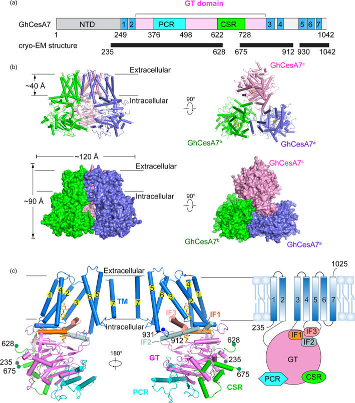
Figure 1.

Cryo‐EM structure of GhCesA7. (a) Schematic of the domains of GhCesA7 (Uniprot: L7NUA2). Seven transmembrane helices are coloured blue. PCR and CSR within the GT domain (pink) are in cyan and green. NTD of GhCesA7 is coloured grey. Black bar labelled with exact residue number indicates the region with a model built by cryo‐EM experimentation. (b) The overall structure of GhCesA7 in side view and top view. Structures are shown as cartoon and surface coloured slate, green, and pink for each protomer. The glucan in each protomer is shown as a stick and coloured yellow. (c) Structure of a GhCesA7 protomer. The transmembrane region is coloured blue, and each helix is numbered. The NTD, PCR, CSR and GT domains are coloured as in (a). The IF1‐3 are coloured orange, pale cyan and salmon, respectively
