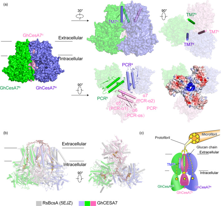
Figure 3.

The trimerization of GhCesA7. (a) The TM7 and PCR of GhCesA7. Side view and top view of TM7 are shown as surface with TM7 in cartoon. The PCR are shown as cartoon and surface. The electrostatic surface potential of PCR was calculated by PyMOL. (b) Structure superimposition of GhCesA7 with RsBcsA (5EJZ). (c) Proposed model of trimeric GhCesA7 assembled for protofibril synthesis. Trimerization of GhCesA produces a protofibril with one glucan chain synthesized by one protomer. The catalytic core is highlighted with a star
