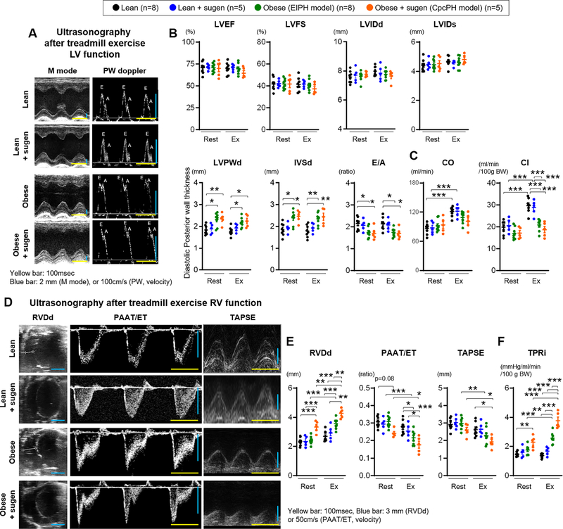
Figure 2. Right ventricular dysfunction after exercise in ZSF-1 obese and obese+sugen models.
(A) Representative images from M mode and pulse-wave (PW) doppler mode ultrasonography. Yellow scale bar, 100 msec. Blue scale bar, 2 mm (M mode), or 100 cm/s (PW mode). (B-C) Left ventricular Ejection fraction (LVEF), Fraction shortening (LVFS), Internal diastolic or systolic diameter (LVIDd or LVIDs), end-diastolic posterior wall (LVPWd), end-diastolic interventricular septal wall thickness (IVSd), E wave/A wave ratio (E/A), cardiac output (CO), and cardiac index (CI) were measured at rest and during exercise. (D) Representative images of M mode and PW doppler mode ultrasonography used to calculate right ventricular end-diastolic diameter (RVDd), pulmonary artery acceleration time (PAAT) per ejection time (ET), and tricuspid annular plane systolic excursion (TAPSE). Yellow scale bar, 100 msec. Blue scale bar, 3 mm (RVDd or TAPSE), or 100 cm/s (PAAT/ET). (E-F) RVDd, PAAT/ET, TAPSE, and TPRi were measured at rest and after exercise. Rats per group; lean n=8, lean+sugen n=5, obese n=8, obese+sugen n=5. Results are expressed as mean±SEM. *P<0.05, **P<0.01, ***P<0.001. Statistical analyses were performed as described in Figure 1 legend.

