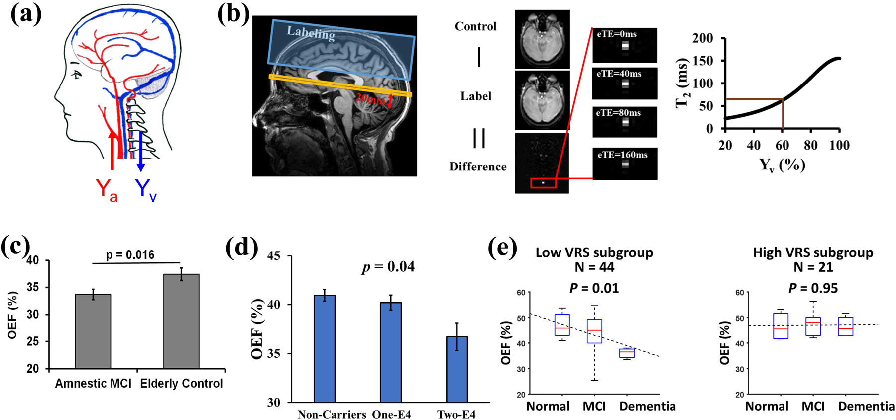
Fig. 4.

Example of OEF determination in neurodegenerative disease. (a) Illustration of arterial (Ya) and venous (Yv) oxygenation fraction in the brain. OEF = (Ya-Yv)/Ya. (b) Measurement of Yv with TRUST MRI. Left panel: Typical positions of imaging slice (yellow) and labeling slab (blue). The red arrow and label indicate that the imaging plane is placed to be parallel to the anterior commissure – posterior commissure (AC-PC) line and 20 mm above the sinus confluence point. Middle panel: Representative raw images of TRUST MRI. Right panel: Conversion from blood T2 to oxygenation fraction. (c) Diminished OEF in patients with mild cognitive impairment (MCI). Reproduced, with permission, from Thomas, B.P. et al. [100]. (d) OEF was decreased in cognitively normal older individuals who have a higher genetic risk (i.e. APOE4) to develop Alzheimer’s disease. Reproduced, with permission, from Lin, Z. et al. [99]. (e) Vascular risk factors (VRS) have an additional effect on OEF. Reproduced, with permission, from Jiang, D. et al. [98].
