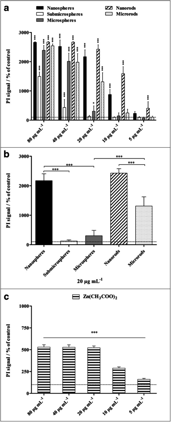
Fig. 8.

Viability of NR8383 alveolar macrophages after 16 h of exposure to different ZnO particles (a, b) and to a zinc acetate solution as control (c). The cell viability was determined by propidium iodide (PI) staining of non-viable cells, and the mean PI fluorescence intensity was assessed by flow cytometry. a Comparison of the effects of different particle concentrations and morphologies. b Comparison of the effects of different particle morphologies at a particle concentration of 20 μg mL−1. All concentrations refer to solid ZnO, except for zinc acetate where the concentration of Zn2+ is given. The data are expressed as mean ± SD (N = 3), given as the percentage of the control (100%, untreated cells). Asterisks (*) indicate significant differences in comparison to the control (*p ≤ 0.05, ***p ≤ 0.001)
