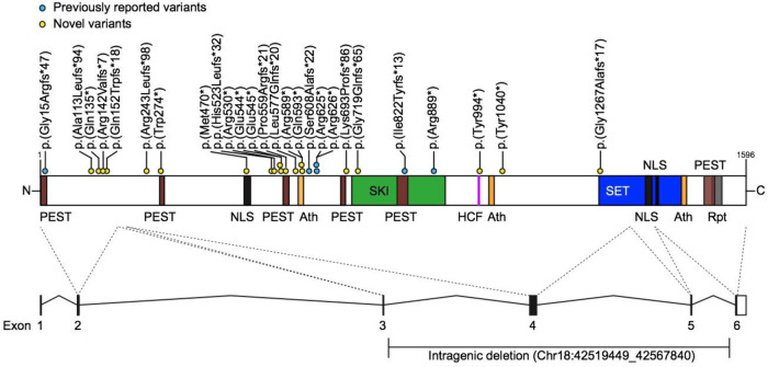
Fig. 1. Location of truncating variants in relation to SETBP1 protein.
Schematic representation of the SETBP1 protein (UniProt: Q9Y6X0) indicating loss-of-function variants included in this study. Five exons (black bars) encode isoform A of the protein (1596 amino acids). Five exons (black bars) encode isoform A of the protein (1596 amino acid residues). The SETBP1 protein sequence contains three AT-hook domains (Ath; orange; amino acids 584–596, 1016–1028, 1451–1463), a SKI homologous region (SKI; green; amino acids 706–917), a HCF1-binding motif (HCF; magenta; amino acids 991–994), a SET-binding domain (SET; blue; amino acids 1292–1488), three bipartite NLS motifs (black; amino acids 462–477, 1370–1384, 1383–1399), six PEST sequences (brown; amino acids 1–13, 269–280, 548–561, 678–689, 806–830, 1502–1526) and a repeat domain (Rpt; grey; amino acids 1520–1543) [31–34]. Blue circles represent previously reported variants and yellow circles indicate novel variants. Two individuals with larger deletions (IND 4, 24) are not shown here. For cDNA annotation of the variants see Table 1.

