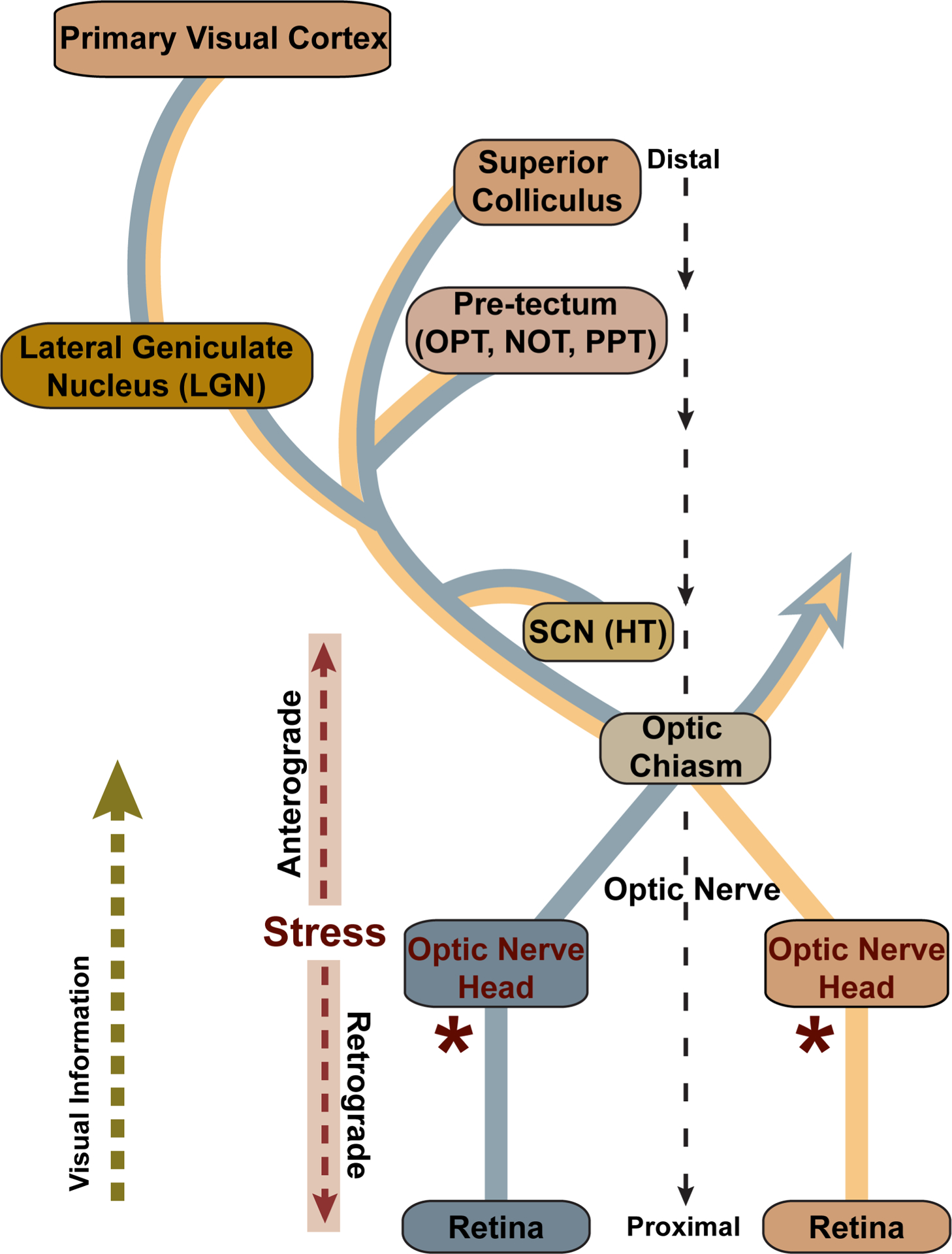
Figure 2. Visual projection in glaucoma.

Retinal ganglion cell axons project through the optic nerve head and at the optic chiasm join either the ipsilateral or contralateral optic tract to central brain targets. The strength of each projection varies by species; the rodent retina projects primarily contralaterally, while the primate retina projects equally to both. Central targets include the suprachiasmatic nucleus (SCN) of the hypothalamus (HT) and the olivary pretectal nucleus (OPT), nucleus of the optic tract (NOT), and posterior pretectal (PPT) nucleus of the pretectum. The lateral geniculate nucleus (LGN) of the thalamus is the primary RGC recipient in primates, while in rodents all or nearly all ganglion cells project to the superior colliculus (SC), while extending axon collaterals to nuclei more proximal to the retina. In all mammals the SC is the most distal direct target, while the LGN provides all direct projection to the primary visual cortex. In the glaucoma, stress at the optic nerve head (*) is conveyed along the ganglion cell axon both towards the brain, in the anterograde direction, and towards the retina, in the retrograde direction. Modified from Crish and Calkins (2015).
