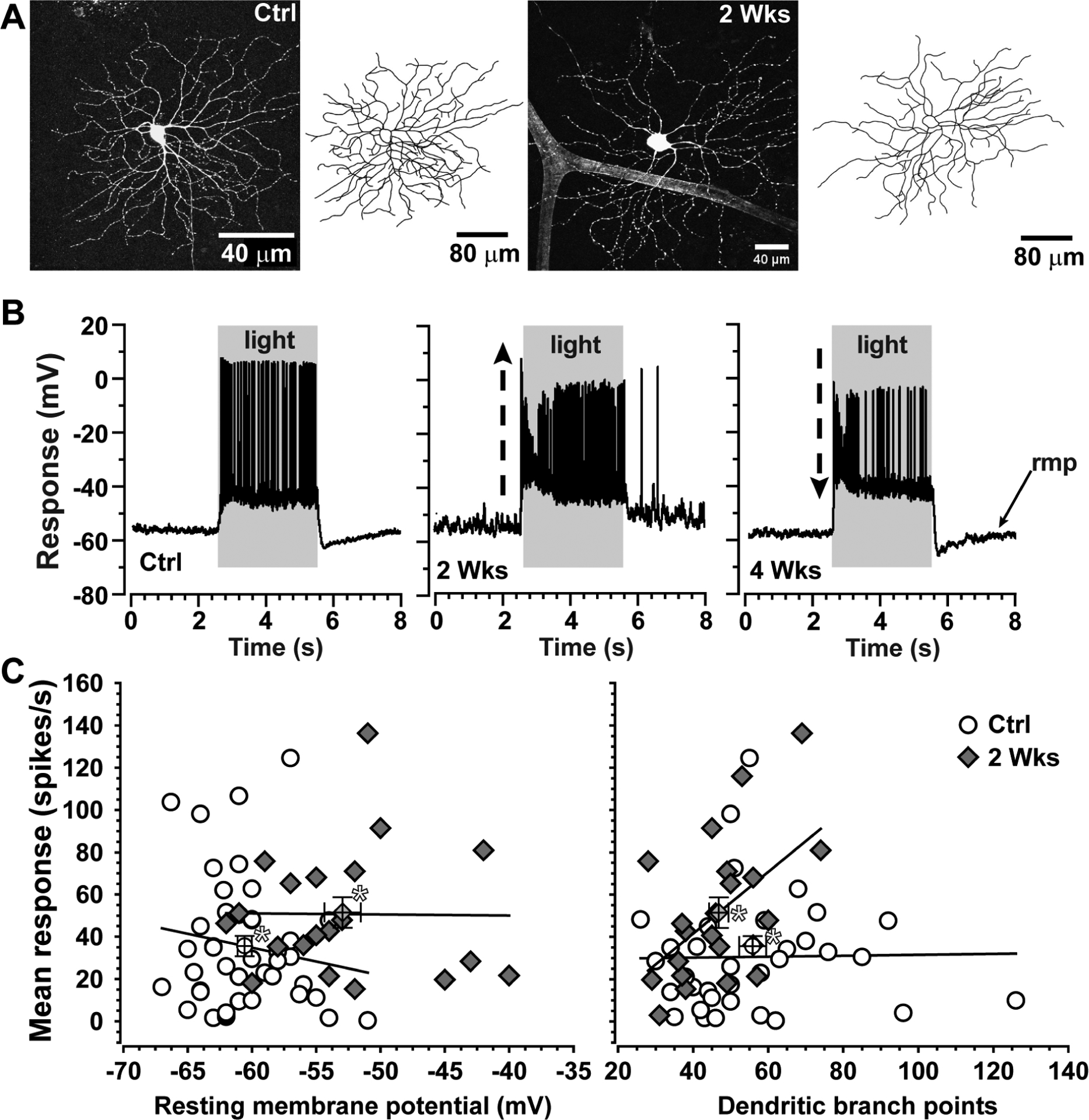
Figure 5. Enhanced excitability is independent of dendritic pruning.

A. Dendritic arbor of an ON-Sustained RGC from control (left) mouse retina following intracellular filling during physiological recording. Same cell type following two weeks of microbead-induced IOP elevation (+35%) shows loss of dendritic branch points (right). B. Current-clamp recordings from single ON-Sustained RGCs from control mouse retina (left) and from retinas following two (middle) and four (right) weeks of elevated IOP. At two weeks, the RGC demonstrates enhanced response to light and a more depolarized resting membrane potential (rmp); by four weeks, response diminishes compared to control. C. Mean response to light after two weeks of elevated IOP increases as resting membrane potential becomes more depolarized (left) and dendritic branch points diminish (right) compared to control (* marks averages ± SEM). Despite having fewer branch points overall, the mean response of ON-Sustained RGCs at two weeks increases with dendritic complexity (p=0.01). Experiments described in Risner et al. (2018).
