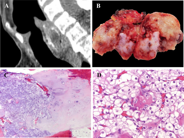
Fig. 1.
a Reformatted sagittal computed tomography image of the larynx demonstrates a focally calcified mass located posteriorly, causing stenosis of the lumen. b Excised chondrosarcoma is solid, lobular, and has an area that is blue-white and regions that are glistening pale tan-yellow. c Low-grade chondrosarcoma arising from chondroma. The chondrosarcoma shows increased cellularity and mild nuclear atypia juxtaposed to the chondroma that is less cellular and lacks atypia. (HES × 10). d Clear cell chondrosarcoma. Sheets of large polygonal tumor cells with abundant clear to pale eosinophilic cytoplasm closely admixed with trabeculae of metaplastic woven bone focally lined by osteoblasts (HES × 40)

