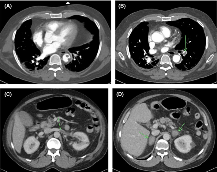
FIGURE 1.

(A) Multiple small filling defects with the descending thoracic aorta adherent to the lateral and posterior wall. (B) Multiple filling defects within the segmental branches of the pulmonary artery of the lower lobe of the left lung. (C) Long filling defect with the left renal vein representing renal vein thrombus, also with wedge shape infarct in left kidney. (D) Bilateral adrenal gland masses with ill‐defined margins representing adrenal hematomas
