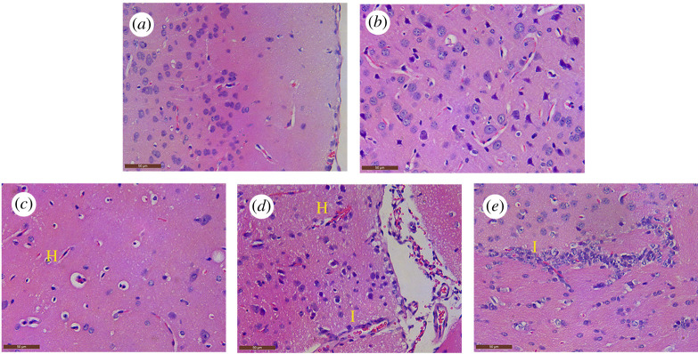
Figure 1.
Histopathological observations of the brain of PRV-inoculated examinated by H&E staining. (a) Microscopic lesion in the control group; (b) 36 hpi group; (c) hyperemia (H, yellow letter) appeared in the brain tissue, followed by perivascular space widened; perivascular lymphocytes increased, and degeneration and necrosis occurred in some neurons in the 48 hpi group; (d,e) focal inflammatory cellular infiltration (I, yellow letter) was found in the brain of the 60 hpi and 72 hpi groups.

