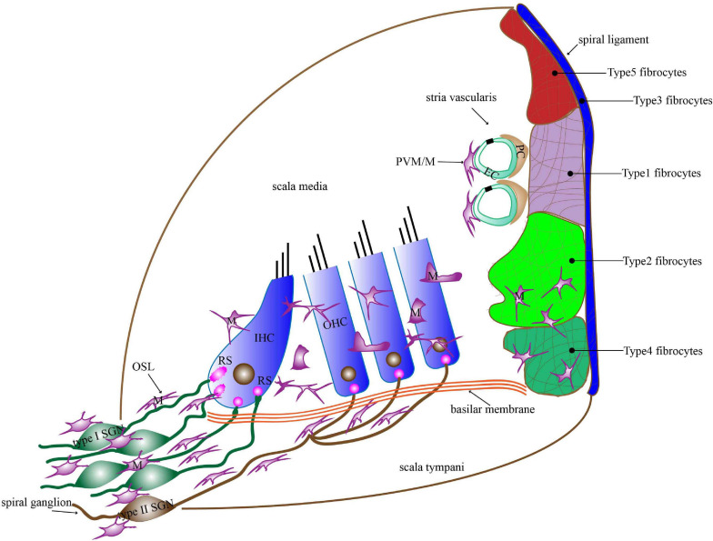
FIGURE 1.
The distribution of macrophages in the basilar membrane, spiral ligament, stria vascularis, OSL, and spiral ganglion. Macrophages in the basilar membrane are distributed below in the HCs on the scala tympani side. Macrophages in the apical turn show dendritic morphology, those in the middle turn display irregular morphology with short projections, and those in the basal turn transform into amoeboid morphology. Macrophages in the lateral wall contain PVM/Ms of the stria vascularis and macrophages among fibrocytes in the spiral ligament. Macrophages are present around the neural tissue, which is composed of SGN cell bodies and their peripheral nerve fibers inside the OSL as well as the modiolus. Most of the macrophages in the OSL area lie in a direction parallel to the radial fibers. M, macrophages; PVM/M, perivascular macrophage-like melanocyte; RS, ribbon synapse; EC, endothelial cells; PC, pericytes; IHC, inner hair cell; OHC, outer hair cell; OSL, osseous spiral lamina; SGN, spiral ganglion neuron.

