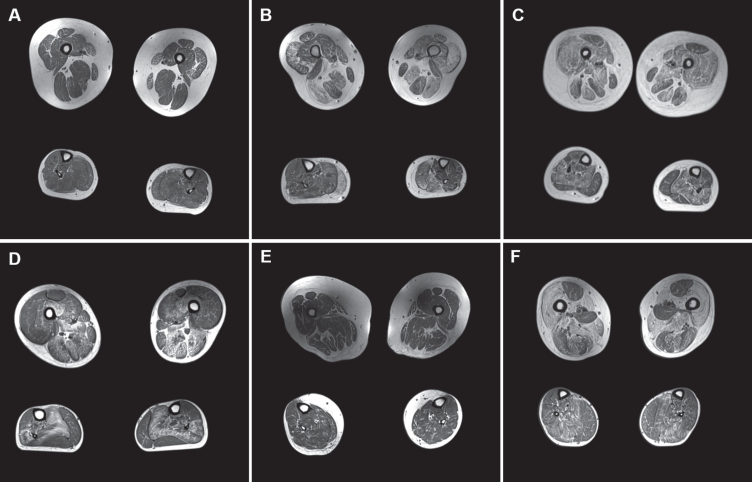
Fig. 1.
A-F: MRI of 6 clinically severe cases. Upper and lower leg slices from cases with clinical severity scores > 5 (A: clinical severity score = 6; B = 5; C = 6; D = 7; E = 6; F = 5) with relative sparing of rectus femoris (in A, E, and F), adductor longus and gracilis in the upper leg, and soleus being the most affected muscle in the lower leg.

