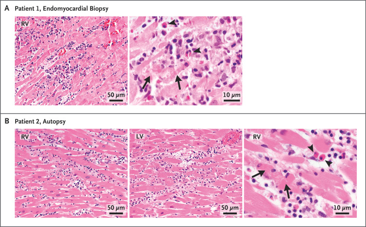
Figure 1. Histopathological Findings from Endomyocardial Biopsy and Autopsy.
Hematoxylin–eosin stains of heart-tissue specimens obtained by means of endomyocardial biopsy in patient 1 (Panel A) and autopsy in patient 2 (Panel B) showed myocarditis in both patients, with multifocal cardiomyocyte damage (arrows) associated with mixed inflammatory infiltration. Scattered eosinophils were noted (arrowheads). The images of the hematoxylin–eosin stains were obtained with 10× eyepieces and 40× or 60× objectives. Additional information is provided in the Supplementary Appendix. RV denotes right ventricle, and LV left ventricle.

