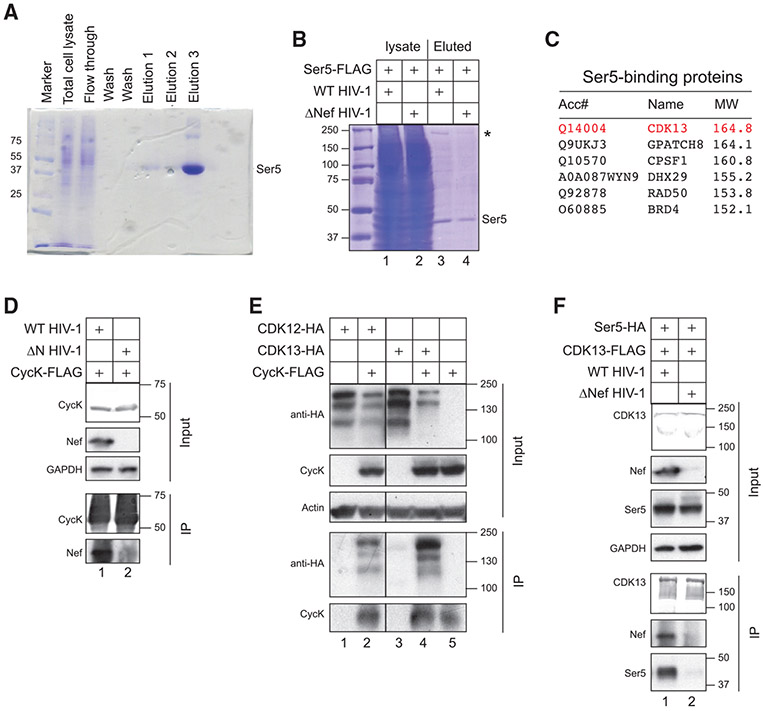
Figure 1. Identification of CycK/CDK13 as a Nef-associated kinase complex via mass spectrometry.
(A) FLAG-tagged SERINC5 protein was expressed in HEK293T cells and purified by anti-FLAG M2 affinity chromatography. After SDS-PAGE, proteins from total cell lysate and three eluted fractions were analyzed after being stained with Coomassie brilliant blue. The monomeric SERINC5 band is labeled.
(B) FLAG-tagged SERINC5 was expressed with a WT or ΔNef HIV-1 proviral vector in HEK293T cells and purified and analyzed similar to as in (A). The monomeric SERINC5 band is labeled and a protein band at ~170 kDa is indicated by an asterisk.
(C) purified proteins from (B) were analyzed by liquid chromatography-mass spectrometry (LC-MS). Control experiments were conducted using beads that were not conjugated with any antibodies. Six proteins with molecular masses of 150–200 kDa that are not found in the control experiments are listed.
(D) FLAG-tagged CycK was expressed with WT or ΔNef HIV-1 in HEK293T cells. Proteins were immunoprecipitated with an anti-FLAG antibody and analyzed by western blotting (WB). CycK was detected by an anti-FLAG antibody and Nef and GAPDH were detected by their specific antibodies. IP, immunoprecipitation; Input, cell lysate.
(E) FLAG-tagged CycK was expressed with HA-tagged CDK12 or CDK13 in HEK293T cells. Proteins were immunoprecipitated and analyzed as in (D). CDK12 and CDK13 were detected by an anti-HA antibody.
(F) FLAG-tagged CDK13 was expressed with HA-tagged SERINC5 in the presence of WT or ΔNef HIV-1 in HEK293T cells. Proteins were immunoprecipitated and analyzed as in (D) and (E).
All experiments were repeated twice, and similar results were obtained.

