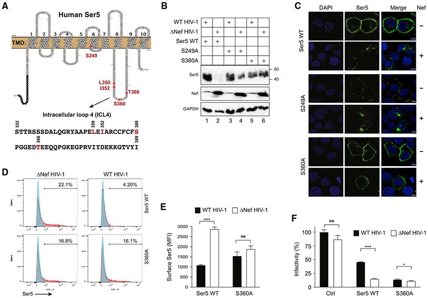
Figure 3. SERINC5 S360 is critical for Nef antagonism of SERINC5.
(A) SERINC5 transmembrane topology and its ICL4 aa sequence are presented. Residues selected for mutagenesis are colored in red.
(B)SERINC5, SERINC5S249A, and SERINC5S360A were expressed with WT or ΔNef HIV-1 in HEK293T cells. Their expression was detected by WB with an anti-FLAG antibody.
(C) SERINC5, SERINC5S249A, and SERINC5S360A with an internal FLAG tag were expressed with WT or ΔNef HIV-1 in HeLa cells. Cells were stained with a fluorescent anti-FLAG antibody, and the antibody uptake was determined by confocal microscopy at 37°C (scale bars, 5 μm).
(D) SERINC5 and SERINC5S360A with an internal HA tag were expressed with WT or ΔNef HIV-1 in HEK293T cells and their surface expression was analyzed by flow cytometry.
(E) MFI values for the SERINC5-positive cell populations in (D) were statistically analyzed.
(F) The anti-HIV-1 activity of SERINC5 and SERINC5S360A was compared and presented similar to as previously.
Error bars in (E) and (F) represent SEM from three independent experiments. Statistical analysis: *p < 0.05, ****p < 0.0001; ns, p > 0.05.

