Fig. 5.

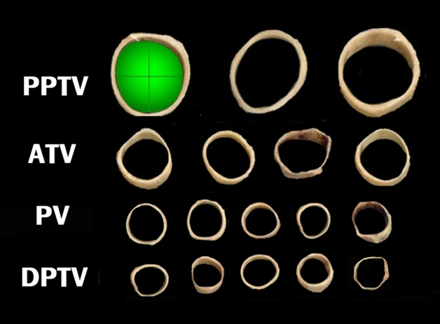
Sliced venous specimens that were scanned and then measured using Image J. The green shaded area represents the surface area, and dotted lines represent the horizontal and longitudinal diameters of the PPTV

Sliced venous specimens that were scanned and then measured using Image J. The green shaded area represents the surface area, and dotted lines represent the horizontal and longitudinal diameters of the PPTV