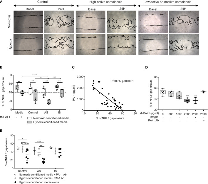
Figure 6.
Secretion of PAI-1 by hypoxic MD-macrophages from high active sarcoidosis inhibited lung fibroblast migration. (A) Representative contrast-phase microscopy images of NHLF during gap closure assays at basal time and after 24hrs of incubation with conditioned media from controls or high active sarcoidosis (AS) or low active or inactive sarcoidosis (IS) patients MD-macrophages exposed to normoxia or hypoxia. (B) Quantitative analysis of NHLF gap closure assay comparing media alone or with 2500pg/ml recombinant human PAI-1 (rh-PAI-1) or normoxic or hypoxic conditioned media from MD-macrophages in controls, AS, and IS. Each point indicates a patient and/or control (n=5-7/group) (C) Correlation (Pearson Test) between PAI-1 level (pg/ml) in normoxic or hypoxic MD-macrophages conditioned media from sarcoidosis and controls and percentage of NHLF gap closure. Each point indicates a patient and/or control (n=3-6/group). (D) Dose effect of rh-PAI-1 on NHLF gap closure reversed by PAI-1 Ab (n=3 independent experiments); (E) Effect of PAI-1 antibody (PAI-1 Ab) added to the conditioned media on NHLF gap closure assay. Each point indicates a patient and/or control (n=3/group). Results are expressed as box plot showing 25th and 75th percentile and median (B, D) or mean with SD (E). *p < 0.05; **p < 0.01; ***p < 0.001; ****p < 0.0001 in Anova two-way (D, E) and Anova two-way with repeated measures (B) with Sidak post-hoc test. #p < 0.05 between normoxic conditioned media from AS and IS.

