Fig. 6.
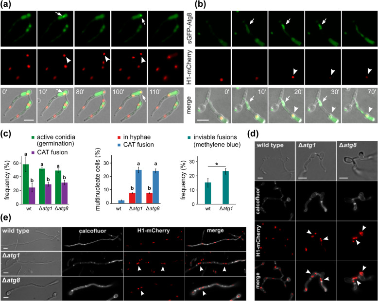
Autophagy is involved in selective nuclear degradation, but not in cell fusion or incompatibility-triggered death. a Live-cell imaging of a self-fusion (Ls.17 H1-mCherry sGFP-Atg8). A ring-like structure with accumulated sGFP-Atg8 (arrows) surrounds the sequestered nucleus (arrowheads). b Live-cell imaging of an “incompatible” fusion (Ls.17 H1-mCherry sGFP-Atg8 × BB H1-sGFP). Localized accumulation of sGFP-Atg8 near the fusion point (arrows) shortly before the incompatibility reaction. Arrowheads: nucleus undergoing degradation. c Frequency of active conidia and their fraction involved in CAT-mediated fusion (left; n = 300 conidia per replicate), non-apical hyphal compartments and fused cells with more than one nuclei (middle; n = 300 hyphal cells or 150 anastomoses per replicate), and inviable fusions determined by staining with methylene blue (right; n = 150 anastomoses per replicate). Wild-type (wt) pairing: Ls.17 H1-mCherry × PH H1-sGFP; pairing of autophagy-deficient mutants (Δatg1): Ls.17 Δatg1 × PH Δatg1. Each strain/pairing was tested in triplicate. Statistical significance of differences was tested with one-way ANOVA followed by Tukey’s post hoc test (left and middle; bars with the same letter do not differ significantly, p value > 0.05) or Student’s t-test (right; * p ≤ 0.05). Error bars: SD. d, e Multinucleate cells (arrowheads) arise frequently in Δatg1 and Δatg8 autophagy-deficient mutants from cell fusion (d) or sub-apical nuclear division (e), in contrast to the wild type that exhibits strictly uninucleate organization. Calcofluor white was used for cell wall staining. Scale bars = 5 μm

