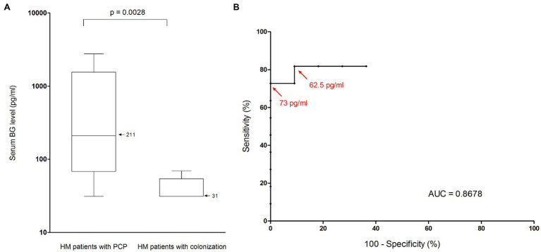
Figure 3.
Performance of BG assay for Pneumocystis pneumonia diagnosis in patients suffering from hematological malignancy. (A) Serum BG levels (pg/ml) in HM patients with Pneumocystis pneupneumonia and in HM patients colonized by Pneumocystis. Black horizontal bars, median values. The median value for HM patients with PCP was 211 pg/ml (range, 31–2,271; interquartile range, 1,489 pg/ml). The median value for HM patients with colonization was 31 pg/ml (range, 31–69; interquartile range, 23 pg/ml). Serum BG levels were significantly lower in the colonization group than in the PCP group (Mann–Whitney test, p = 0.0028). (B) Receiver operating characteristic curve for serum BG assay performance in HM patients, using microscopic detection of P. jirovecii as the reference method. Red arrows represent calculated thresholds for maximal specificity (73 pg/ml) and maximal sensitivity (62.5 pg/ml). AUC, area under the curve; BG, (1,3)-β-D-glucan; HM, hematological malignancy; and PCP, Pneumocystis pneumonia.

