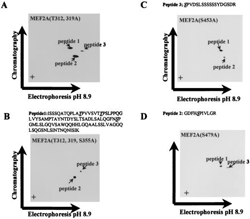
FIG. 5.
In vitro phosphorylation sites of MEF2A by p38. (A) Phosphopeptide mapping of MEF2A(T312, 319A) phosphorylated by p38 in vitro; (B) phosphopeptide mapping of MEF2A(T312, 319, S355A) phosphorylated by p38 in vitro; (C) phosphopeptide mapping of MEF2A(S479A) phosphorylated by p38 in vitro; (D) phosphopeptide mapping of MEF2A(S453A) phosphorylated by p38 in vitro. The sequences of predicted phosphopeptides are shown above panels B to D, with phosphorylated residues underlined.

