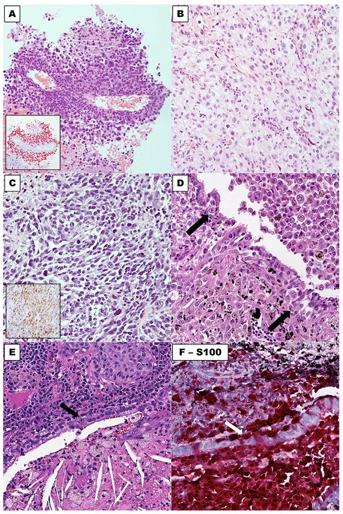
Fig. 2. Microscopic features of lung-only melanomas.

Majority of cases show predominant epithelioid cytomorphology, which may mimic non-small cell carcinoma (a, b), and some cases have spindle cell morphology (c). Expression of SOX10 and HMB45 is illustrated in insets in (a) and (c), respectively. An example (d) showing pigmented melanoma cells involving the bronchial epithelium in small nests (arrow: melanoma cells in bronchial epithelium). e and f illustrate H&E stain and S100, respectively, of involvement of bronchial epithelium adjacent to a lung-only melanoma. Arrows: melanoma cells in bronchial epithelium.
