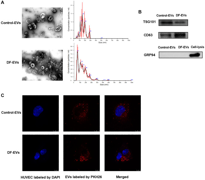
Figure 1.
Characteristics of EVs derived from different wound fluids. (A) TEM analysis and NTA analysis of EVs. Scale bar: 200 nm. (B) EVs-related markers were detected by Western blotting. (C) EVs were labeled with PKH26 and co-incubated with HUVECs, and the representative images photographed by confocal microscope are showed above. Scale bar: 10 μm. At least three replicates of each experiment were performed.

