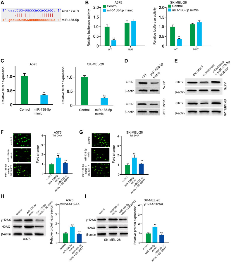
Figure 5.
MiR-138-5p induces DNA damage by targeting SIRT7 in melanoma cells. (A) The binding of miR-138-5p and SIRT7 3’UTR was identified by bioinformatic analysis based on Targetscan (http://www.targetscan.org/vert_72/). (B–D) The A375 and SK-MEL-28 cells were treated with control mimic or miR-138-5p mimic. (B) The luciferase activities of wild type SIRT7 (SIRT7 WT) and SIRT7 with the miR-138-5p-binding site mutant (SIRT7 MUT) were examined by luciferase reporter gene assays in the cells. (C) The mRNA expression of SIRT7 was analyzed by qPCR assays in the cells. (D) The protein expression of SIRT7 was determined by Western blot analysis in the cells. (E) The A375 and SK-MEL-28 cells were treated with control shRNA or circZNF609 shRNA, or co-treated with circZNF609 shRNA and miR-138-5p inhibitor. The protein expression of SIRT7 was tested by Western blot analysis in the cells. (F–I) The A375 and SK-MEL-28 cells were treated with the control mimic or miR-138-5p mimic, or co-treated with miR-138-5p mimic and pcDNA3.1-SIRT7. (F and G) The DNA damage was analyzed by comet assays in the cells. (H and I) The protein expression of H2AX, γH2AX and β-actin was determined by Western blot analysis in the cells. The results of Western blot analysis were quantified by ImageJ software. Data are presented as mean ± SD. Statistic significant differences were indicated: *P < 0.05, **P < 0.01.

