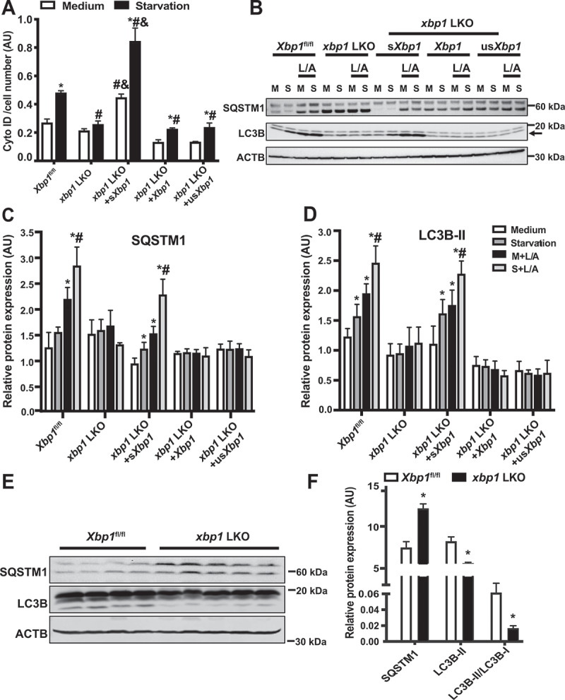
Figure 4.

sXBP1 regulates hepatic autophagy. (A) Intensity of autophagosomotropic dye (Cyto-ID) in live primary hepatocytes from Xbp1fl/fl mice and xbp1 LKO mice transduced with the indicated adenoviral constructs, the Ad-GFP was used as a control virus. Cells were isolated then subsequently treated with EBSS (starvation, 4 h). * indicates statistical significance compared to treatment with the medium in cells harboring the same construct, # indicates statistical significance compared to the Xbp1fl/fl group with the same treatment, and & indicates statistical significance between xbp1 LKO groups with the same treatment (n = 4, biological replicates). (B) Representative western blots of converted LC3B (arrow marks LC3-II) and SQSTM1 protein in primary hepatocytes from Xbp1fl/fl mice and xbp1 LKO mice as in (A). L/A: ammonium chloride and leupeptin, 4 h. Each lane contains protein lysate from an individual mouse. (C and D) Densitometric analysis of western blot in (B). The data were normalized to those of ACTB. * indicates statistical significance compared to the medium group in the same type of cells, and # indicates statistical significance between starvation groups in the same type of cells (n = 3 biological replicates). (E) Representative western blots and densitometric analysis (F) of LC3B and SQSTM1 in livers from Xbp1fl/fl and xbp1 LKO mice under fasting (16 h) conditions. The data were normalized to ACTB. *indicates statistical significance compared to the Xbp1fl/fl group (n = 4–5 biological replicates). All data are presented as means ± SEM. Statistical significance was determined by ANOVA followed by Tukey’s multiple comparisons test in A, C and D, and Student’s t-test in F, p < 0.05
