
Figure 2.
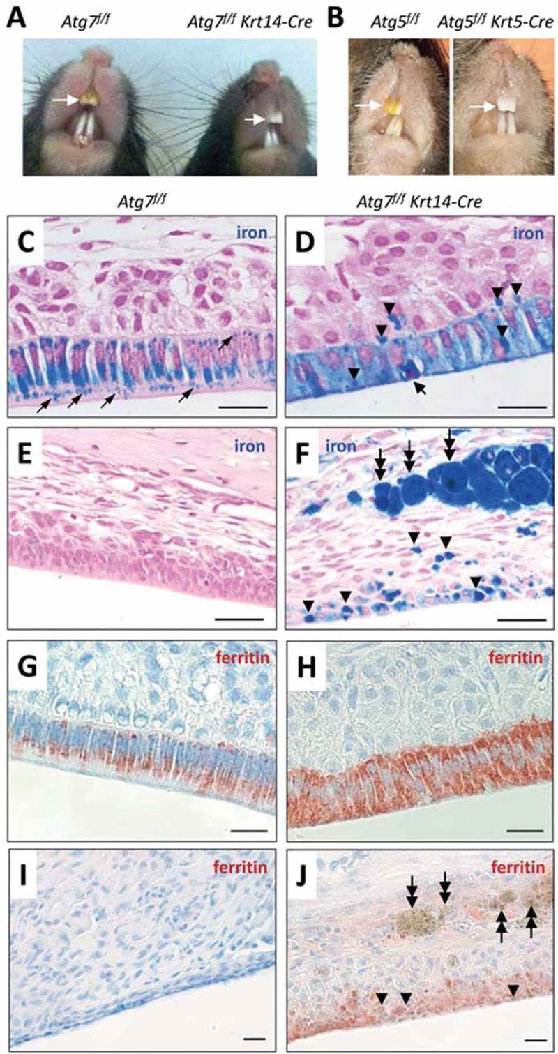
Ameloblasts of Atg7f/f Krt14-Cre mice have a defect in iron secretion. (A) Comparison of incisor colors of an Atg7f/f (fully autophagy-competent) and an Atg7f/f Krt14-Cre (epithelial autophagy-deficient) mouse. (B) Comparison of incisors of an Atg5f/f and an Atg5f/f Krt5-Cre mouse. The maxillary incisors are marked by white arrows (A, B). (C-J) Sections through the enamel epithelium were stained with the iron-specific dye Perls’ Prussian blue and counterstained with nuclear fast red (C-J) or immunostained for FTH1 (red) and counterstained with hematoxylin (blue) (G-J). Images show the pigmentation stage epithelium (C, D, G, H) and the distal epithelium (E, F, I, J). Thin arrows (C) indicate iron-containing vesicles. A thick arrow (D) indicates a condensed nucleus of an apoptotic ameloblast. Arrowheads (D, F, J) indicate accumulations of iron, and two-headed arrows (F, J) indicate iron-containing macrophages. Scale bars: 20 µm (C-J).