
Figure 3.
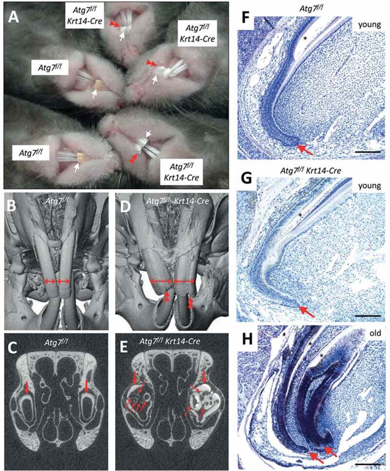
Inactivation of Atg7 in epithelial cells leads to the development of malformations during the growth of maxillary incisors in Atg7f/f Krt14-Cre mice. (A) Comparison of incisors of Atg7f/f and Atg7f/f Krt14-Cre mice aged over 1.5 years. The maxillary incisors are marked by white arrows. Red double arrowheads indicate malformations and damage of incisors of old Atg7f/f Krt14-Cre mice. (B-E) The heads of old Atg7f/f (24 months) and Atg7f/f Krt14-Cre (22 months) mice were investigated by micro-computed tomography. Front view of Atg7f/f (B) and Atg7f/f Krt14-Cre (D) heads showed an aberrant increase in the width of maxillary incisors (horizontal double-headed arrows) and irregular damage (double arrowheads) at the apical ends of Atg7f/f Krt14-Cre incisors. Frontal sections of micro-CT revealed that the incisors of Atg7f/f mice (C) consisted of a single tube (thick arrow), representing the normal condition, whereas incisors of Atg7f/f Krt14-Cre mice (E) contained additional incisor tubes (thin arrows). Micro-CT data are representative for n = 3 mice of each genotype. (F-H) Mice of different age (young: 1–2 months, old: 22 months) were sacrificed, the heads were fixed in formaldehyde, de-calcified and sagittal-sectioned. The tissue sections were subjected to hematoxylin and eosin staining. Young Atg7f/f (F) and Atg7f/f Krt14-Cre (G) mice contained a single cervical loop (arrow) from which a normally shaped maxillary incisor grew. In old Atg7f/f Krt14-Cre (H) an additional cervical loop had formed, giving rise to an ectopic incisor. Scale bars: 200 µm.