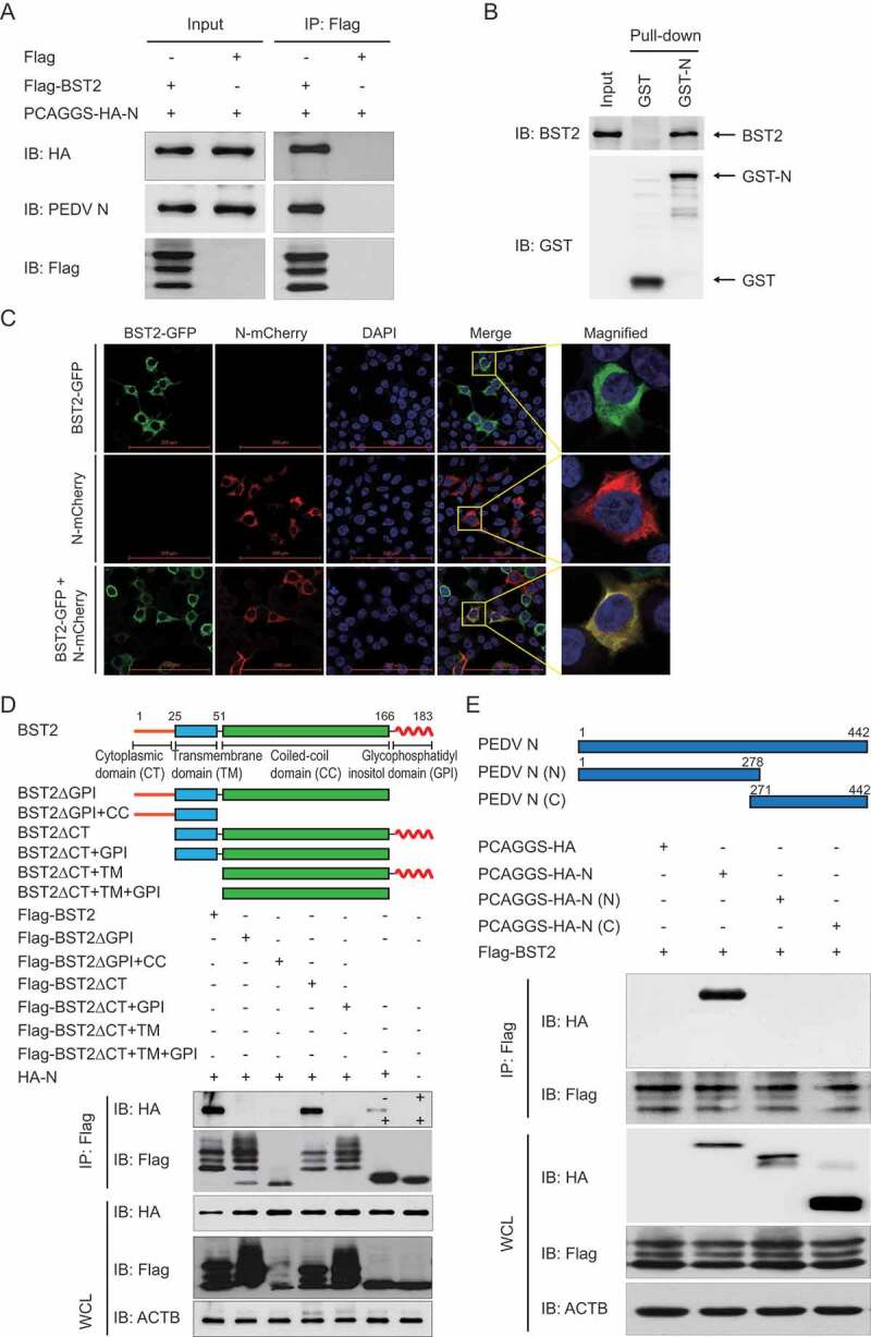
Figure 4.

BST2 interacts with PEDV N protein. (A) Vero cells were transfected with plasmids encoding HA-N and Flag-BST2 or empty vectors for 24 h, followed by Co-IP with anti-Flag binding beads and a western blotting analysis with anti-HA, anti-PEDV N, and anti-Flag antibodies. (B) The full length of the BST2 gene and PEDV N gene were cloned into pCold TF plasmid and pCold GST plasmid, respectively. Recombinant proteins were expressed in bacterial strain BL21 (DE3) and purified for the GST pull-down analysis. After adequate washing, proteins eluted from beads were analyzed by western blotting. Input, BST2. (C) 293T cells were transfected with plasmids encoding BST2-GFP and N-mCherry for 24 h. Cell nuclei were labeled with DAPI, and the fluorescent signals were observed with confocal immunofluorescence microscopy. 293T cells were transfected with plasmids encoding BST2-GFP or N-mCherry as control. Scale bars: 100 μm. (D) 293T cells were transfected with plasmids encoding HA-N and Flag-BST2 or the indicated BST2 mutants. They were then analyzed with Co-IP with anti-Flag binding beads and western blotting with anti-HA and anti-Flag antibodies. Throughout was the western blot analysis of whole-cell lysates (WCLs) without immunoprecipitation. ACTB was used as the sample loading control. (E) Co-IP and western blotting analyses of 293T cells transfected with PEDV N or the indicated N mutants, together with a vector encoding Flag-BST2.
