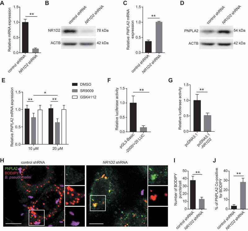
Figure 6.

NR1D2 mediates lipid accumulation through inhibiting PNPLA2 expression following B. pseudomallei infection. (A and B) The mRNA and protein levels of NR1D2 were measured in A549 cells after transfection with control or NR1D2 shRNA (1 μg) for 24 h. (C and D) The mRNA and protein levels of PNPLA2 in control and NR1D2-silenced A549 cells after infected with B. pseudomallei (MOI = 10) for 12 h. (E) The expression of PNPLA2 was measured in A549 cells. After pretreatment with SR9009 or GSK4112 in different concentrations (10 μM and 20 μM) for 48 h, cells were infected with B. pseudomallei (MOI = 10) for 12 h. (F) A549 cells were transfected with either pGL3-Basic empty vector (200 ng) or −2000/+20-LUC PNPLA2 construct (200 ng) plus pRL-TK-Renilla (10 ng) and then infected with B. pseudomallei (MOI = 10) for 12 h. Luciferase activities were measured and normalized to Renilla internal control. (G) −2000/+20 LUC PNPLA2 (200 ng) plus pRL-TK-Renilla (10 ng) was transfected into A549 cells with control or pcDNA3.1-NR1D2 (200 ng) for 24 h. (H) Representative images of control and NR1D2-silenced A549 cells infected with B. pseudomallei for 12 h. Scale bar: 10 μm. (I and J) Quantitative analysis of the number of lipid droplets and the colocalization of PNPLA2 and BODIPY shown in (H). Data is shown as the mean ± SD of three independent experiments. *P < 0.05, **P < 0.01
