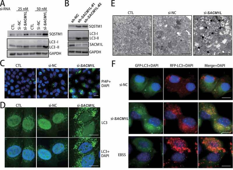
Figure 6.

Human SACM1L functioned in autophagy through regulating autophagosome fusion with lysosomes. (A and B) Knockdown of human SACM1L in HeLa cells resulted in accumulation of SQSTM1 and LC3. siRNA- or shRNA-targeting human SACM1L were transfected into HeLa cells for 48 h and protein levels of autophagic substrate SQSTM1 and autophagosome marker LC3 (human homolog of yeast Atg8) were detected with specific antibodies. siRNA or shRNA with scrambled sequence transfected cells (NC) or no transfection cells (CTL) were used as negative controls. (C) SACM1L deficiency caused a dramatic increase of PtdIns4P levels in HeLa cells. HeLa cells were transfected with siRNAs indicated for 48 h. PtdIns4P-specific antibodies were used for immuno-fluorescence observation. Experiments were conducted for at least three times and representative images were shown. Scale bars: 10 μm. (D) Knockdown of human SACM1L caused accumulated autophagosomes in HeLa cells. HeLa cells were transfected with siRNAs indicated for 48 h and LC3 dots (representing autophagosomes) were observed through immunofluorescence observation. Representative images were shown. Scale bars: 10 μm. (E) Knockdown of human SACM1L caused accumulated autophagosomes in HeLa cells. Control cells and SACM1L knockdown cells were analyzed by transmission electron microscopy. Arrows point to the accumulated autophagosomes in cells. Representative images from three replicated experiments were shown. Scale bars: 0.5 μm. (F) Knockdown of human SACM1L blocked autophagosome fusion with lysosomes in HeLa cells. GFP-RFP-LC3-expressing HeLa cells were transfected with indicated siRNAs or subject to starvation with EBSS media. Autophagosome marker LC3 was monitored by fluorescence microscope. Higher level of RFP-LC3 signal and lower level of GFP-LC3 signal indicated the stimulated autophagy flux while the similar signal levels of RFP-LC3 and GFP-L3 (showing yellow in merge) indicated the blocked fusion step of autophagy flux. Representative images were shown. Scale bars: 10 μm
