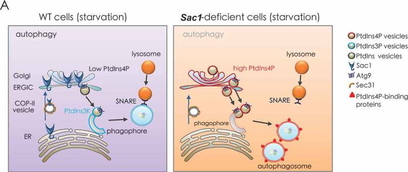
Figure 7.

A proposed model for Sac1 function in autophagy through restraining PtdIns4P from incorporation into autophagosomes. Left, when wild type cells were under starvation conditions, Sac1 trafficked to Golgi by COP-II vesicles to hydrolyze PtdIns4P there. Then the Golgi sourced lipid vesicles with low levels of PtdIns4P were incorporated into autophagosomes. SNARE complex was next recruited to autophagosomes for their fusion with lysosomes. Right, when Sac1 was depleted, the higher level of PtdIns4P in the Golgi led to abnormal integration of PtdIns4P-containing vesicles into autophagosomes. The PtdIns4P enriched autophagosomes s failed to recruit SNARE complex and thus could not fuse with lysosomes
