
Figure 4.
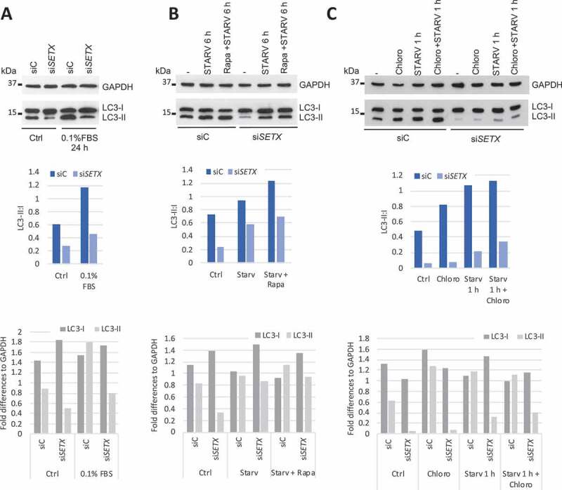
The autophagy flux is defective in SETX-depleted cells. (A) U87 cells were transfected with siC and siSETX for 3 d and grown in normal (Ctrl) or starvation condition for 24 h and 0.1% FBS. LC3 level (LC3-I and LC3-II) was analyzed by WB. GAPDH is used as loading control. (B) U87 cells were transfected as in A and grown under normal conditions (-), starvation in medium without FBS for 6 h (STARV) or in combination with rapamycin at 10 nM for 6 h (Rapa + STARV). (C) U87 cells were transfected as in A and grown under normal conditions (-), treated with chloroquine at 20 μM for 1 h (Chloro), starved in medium without FBS for 1 h or starved in combination with chloroquine treatment for 1 h (Chloro + STARV). Graphs show the quantifications of LC3-II:LC3-I and LC3-I and LC3-II levels normalized to GAPDH