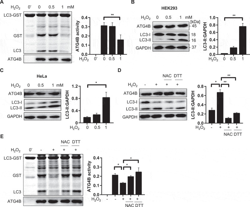
Figure 2.

Endogenous ATG4B activity is inhibited by H2O2. (A) HEK293 cells were treated with designated concentration of H2O2 for 1 h, then cells were harvested in non-denatured buffer. Cell lysate (8 µg) was incubated with substrate LC3-GST (3 µg) at 37℃ for 30 min. The cleavage of LC3-GST by ATG4B was presented by CBB staining and relevant expression level of ATG4B was analyzed by immunoblot with anti-ATG4B antibody. (B-C) Deconjugation activity of endogenous ATG4B in vivo. HEK293 (B) and HeLa (C) cells were exposed to indicate H2O2 for 1 h, respectively. Relevant LC3-II level was detected by immunoblot analysis with anti-LC3B antibody. (D) HeLa cells pretreated with NAC (10 mM) for 15 min were incubated with H2O2 (1 mM) for 1 h, or DTT (1 mM) was added for 15 min after cells were exposed to H2O2 for 1 h. LC3-II level was determined as described in (B-C). (E) Cell lysates (8 µg) from cells processed in (D) were utilized to test processing activity toward LC3-GST (3 µg), followed by CBB staining. Graphical data are presented as mean ± SEM from 3 individual experiments. * P < 0. 05; ** P < 0.01.
