
Figure 5.
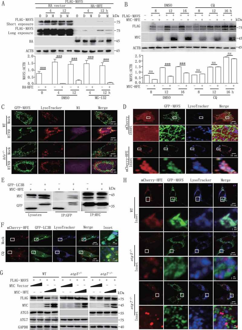
HFE acts as a novel suppressor of MAVS stability through the autophagy-lysosome pathway. (A and B) HEK293T cells were transfected with FLAG-MAVS and HA-HFE plasmids for 24 h and then treated with 20 μM MG132 (M) (A) or 50 µM CQ (B) for the indicated times. The total cell lysates were immunoblotted with the indicated antibodies. (C) Confocal microscopy of WT and hfe−/- BMDMs transfected with GFP-Mavs (green) and stained with LysoTracker Red DND-99 (red) after H7N9 virus infection for 18 h. Scale bars, 10 μm. (D) Confocal microscopyof hfe−/- BMDMs transfected with mCherry-Hfe (red) and GFP-Mavs (green) for 24 h and then stained with LysoTracker Deep Red (blue) for 2 h. Scale bars: 10 μm (1 μm in the inset). (E) MYC-HFE and GFP-LC3B were co-transfected into HEK293T cells. Immunoprecipitation and immunoblotting analysis of the lysates were performed with various combinations of anti-MYC anti-FLAG and anti-GFP antibodies. (F) hfe−/- BMDMs transfected with the mCherry-Hfe (red) expression vector and GFP-Lc3b (green). These BMDMs were stained with LysoTracker Deep Red (blue) for 2 h after treated with 50 μM CQ for another 12 h. Scale bars: 10 μm (1 μm in the inset). (G) Immunoblotting analysis of FLAG-MAVS in lysates of WT, atg5−/- and atg7 −/- cells transfected with different doses of MYC-Hfe. The total cell lysates were immunoblotted with the indicated antibodies. (H) WT, atg5−/- and atg7 −/- MEFs were co-transfected with GFP-Mavs (green) and mCherry-Hfe (red). Cells were stained 24 h later with LysoTracker Deep Red (blue) for 2 h and subjected to confocal microscopy. Scale bars: 10 μm (1 μm in inset). Data are representative of at least three independent experiments, and differences between the experimental and control groups are determined by 2-way ANOVA (***p < 0.001)