Figure 6.

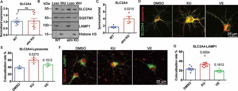
SLC2A4 trafficking switched to lysosomes in the absence of ATM. (A) Normalized Slc2a4 mRNA expression level in WT and atm−/− (atm KO) mouse cortex. Error bars = SEM; n = 8 animals each group; ns, p = 0.8746 (unpaired t test). (B) Representative SLC2A4, SQSTM1, LAMP1 and Histone H3 blots in isolated lysosomes (Lyso) or whole-cell lysates (Wcl) from WT or atm−/− mouse cortex lysates. (C) The ratio of lysosomal SLC2A4 to total SLC2A4 in WT and atm−/− mouse cortex. Error bars = SEM; n = 3–4 animals each group; p = 0.0215 (unpaired t test). (D) Representative images of WT cortical neurons transfected with N1-Slc2a4-GFP and mCherry-Lysosome-20 treated with DMSO, KU-60019 (1 µM) or VE-822 (0.2 µM) for 24 h. (E) Colocalization rate of GFP-SLC2A4 and the mCherry-lysosome. Error bars = SEM; n = 3 batches of cultures; p values are as indicated (unpaired t test). (F) Representative co-immunostaining images of SLC2A4 (red) and LAMP1 (green) in WT cortical neurons treated with DMSO, KU-60019 (1 µM) or VE-822 (0.2 µM) for 24 h. (G) Colocalization rate of endogenous SLC2A4 and LAMP1. Error bars = SEM; n = 10–11 coverslips from 3 batches of cultures; p values are as indicated (unpaired t test)
