FIG. 1.
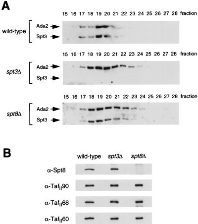
Western blot characterization of Superose 6-purified spt3Δ and spt8Δ SAGA complexes. (A) SAGA complexes derived from wild-type (FY631), spt3Δ (FY294), and spt8Δ (FY462) strains were pooled, concentrated, and run on a Superose 6 column for separation based on molecular weight. Western blots of these fractions were performed to compare the sizes of the complexes. Blots were visualized with dilutions of antisera raised against the Ada2 or Spt3 proteins as indicated. (B) Western blots of wild-type (fraction 20), spt3Δ (fraction 21), and spt8Δ (fraction 21) SAGA, visualized with antisera specific to Spt8, TafII90, TafII68, or TafII60.

