
Figure 3.
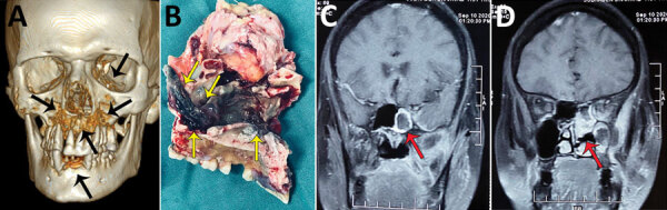
Radiographic images and surgical specimens demonstrating rhino-orbital-cerebral coronavirus disease–associated mucormycosis in patients from India, 2020. A) Three-dimensional reconstruction of computed tomography scan of 54-year-old male patient. Black arrows indicate patchy osteonecrosis involving the upper jaw, right orbital wall, and paranasal sinuses. B) Surgical specimen from the maxilla of 54-year-old male patient showing black necrotic paranasal sinus with palatal involvement indicated by yellow arrows. C, D) Magnetic resonance imaging (MRI) of coronal section of paranasal sinus and brain of 51-year-old female patient. Red arrow in panel C indicates enhancing cavernous sinus lesion; D) red arrow in panel D indicates right ethmoid and maxillary sinusitis. Scale bar indicates 7 cm.