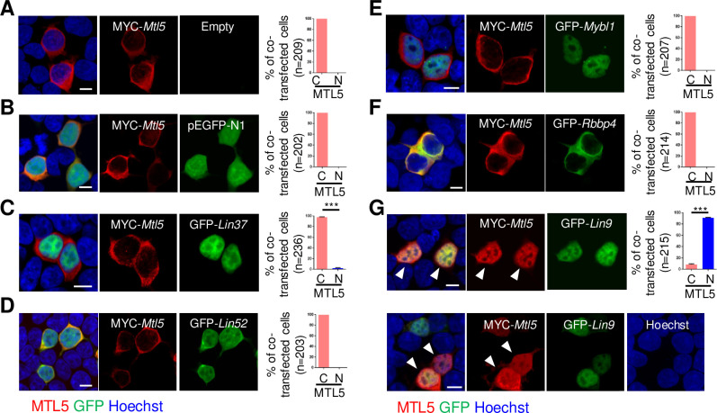
Fig 3. MTL5 is transported into the nuclei of cultured HEK293T cells by LIN9.
HEK293T cells were transiently transfected with plasmids encoding (A) MYC-Mtl5, (B) MYC-Mtl5 and pEGFP-N1, (C) MYC-Mtl5 and GFP-Lin37, (D) MYC-Mtl5 and GFP-Lin52, (E) MYC-Mtl5 and GFP-Mybl1, (F) MYC-Mtl5 and GFP-Rbbp4, or (G) MYC-Mtl5 and GFP-Lin9. The localization of proteins are observed by immunostaining with antibodies against MTL5 (red) and GFP (green) antibodies, and imaged by a Nikon confocal laser scanning microscope system. Whit arrowheads indicate the co-localization of MTL5 and LIN9. The cells with nuclear (N) or cytoplasmic (C) localization of MTL5 were counted and “n” in the brackets represents the number of co-transfected cells that were analyzed. Data are presented as mean ± SEM from three independent experiments. P values were analyzed by t-test. ***p<0.001. Scale bars, 10 μm.

