
Fig. 4. Outward open conformation of PepT2.
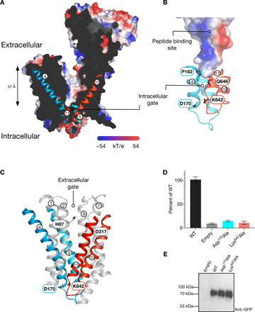
(A) Slice through the electrostatic surface representation of PepT2 as viewed in the plane of the membrane. The intracellular gating helices TM4 to TM5 and TM10 to TM11 are shown. (B) Zoomed-in view of the intracellular gate, with the key stabilizing interactions identified and labeled. (C) Cartoon representation showing the gating helices in PepT2. Key side chains involved in gate stabilization are shown. (D) Cell-based transport assays for PepT2 and variants of the intracellular gate–interacting side chains. (E) Western blot using an anti–green fluorescent protein (GFP) antibody to detect WT and variant forms of PepT2.