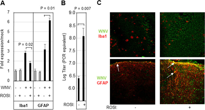
FIG 2.
Rosiglitazone inhibits microglial activation and leads to increased viral titer in WNV-infected BSC. BSC were infected with WNV (105 PFU per slice) in the presence/absence of 20 μM rosiglitazone (ROSI). (A to C) Five days following infection, cultures were harvested, and RNA was prepared. WNV-induced expression of microglial (Iba1) and astrocyte (GFAP) markers (A, average increased expression over mock), as well as viral titer (B, average titer) is shown. Error bars indicate the standard error of the mean (n = 3). Statistical significance was determined using 2-sample, 2-tailed t tests (GraphPad). (C) WNV-infected slices were also examined by immunohistochemistry. The images show decreased Iba1 staining (red staining in upper images) in rosiglitazone-treated, WNV-infected slices compared to untreated, infected controls. In contrast, rosiglitazone treatment of WNV-infected BSC resulted in increased GFAP expression (red staining in lower images) compared to untreated, infected controls. Counterstaining of slices with WNV antibody (green staining) revealed increased viral antigen in rosiglitazone slices compared to untreated, WNV-infected controls. Cells that stained yellow (white arrows) are positive for both GFAP and WNV. The images shown are representative of 3 individual slices.

