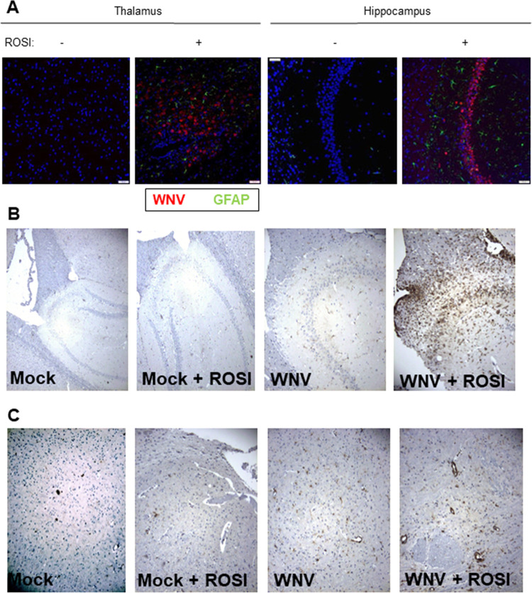
FIG 5.
Rosiglitazone increases the activation of astrocytes, viral growth, and lymphocyte infiltration in the brains of WNV-infected mice. Mice were fed rosiglitazone chow for 14 days prior to footpad inoculation of 1000 PFU WNV (NY99). At 9 days postinfection, brains were harvested, sectioned, and stained with antibodies directed against GFAP (A), WNVE (A), or CD3 (B and C). Similar to what we observed in ex vivo slices, and consistent with data presented in Fig. 4, rosiglitazone treatment resulted in higher levels of WNV antigen (red staining) (A), GFAP (green staining) (A), and CD3 (B and C) in the thalamus (A), hippocampus (A and B), and cortex (C) of the WNV-infected/rosiglitazone-treated, WNV-infected mouse with the highest caspase 3 positivity and greatest level of injury compared to an untreated, WNV-infected, control. Increased CD3 staining is also seen in the hippocampus (B) and cortex (C) of an untreated, WNV-infected mouse compared to mock-infected controls.

