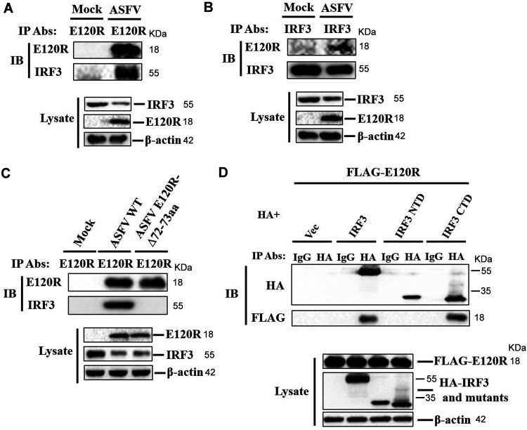
FIG 8.
E120R interacted with IRF3 during viral infection. (A) PAMs were mock infected or infected with ASFV (MOI of 1) for 12 h. The cell lysates were immunoprecipitated with anti-E120R antibody and subjected to Western blotting using the indicated antibodies. Three independent experiments were performed. IP, immunoprecipitation; IB, immunoblotting. (B) A reverse immunoprecipitation assay was performed using anti-IRF3 antibody as described in the legend to panel A. The antibody-antigen complexes were detected using anti-E120R or anti-IRF3 antibody. Three independent experiments were performed. (C) PAMs were mock infected or infected with ASFV WT or ASFV E120R-Δ72-73aa (MOI of 1) for 12 h. The cell lysates were immunoprecipitated with anti-E120R antibody and subjected to Western blotting using the indicated antibodies. Three independent experiments were performed. (D) HEK-293T cells were transfected with 8 μg of FLAG-E120R expression plasmid along with 8 μg of HA vector or HA-IRF3, HA-IRF3 NTD, or HA-IRF3 CTD expression plasmid for 24 h. Cells were lysed, and the lysates were immunoprecipitated with anti-HA antibody and subjected to Western blotting. NTD, N-terminal domain; CTD, C-terminal domain. Three independent experiments were performed.

