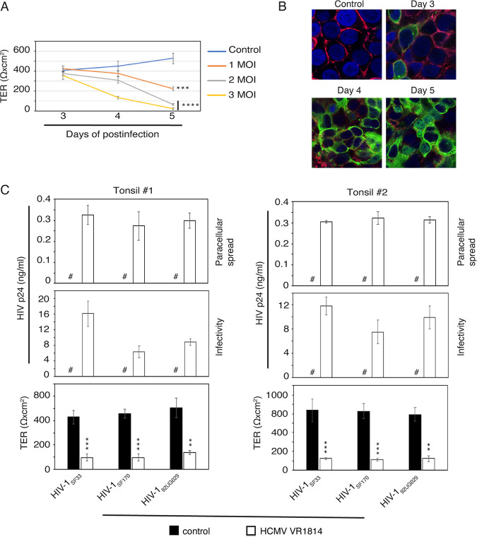
FIG 7.
HCMV infection disrupts tonsil epithelial junctions and facilitates paracellular HIV-1 spread. (A and B) Polarized tonsil epithelial cells were infected with 1, 2, or 3 MOI of HCMV VR1814, and TER was measured after 3, 4 and 5 days (A). One set of cells infected with HCMV VR1814 at an MOI of 3 was coimmunostained for occludin (red) and HCMV gB (green) after 3, 4, or 5 days (B). Uninfected polarized cells served as a control. Cell nuclei were stained with DAPI (blue), and cells were analyzed by fluorescence microscopy. Magnification, ×400. (C) Polarized tonsil epithelial cells from two independent donors were infected with HCMV VR1814 for 5 days. TER was measured (bottom), and the apical surface of cells was exposed for 2 h to dually tropic HIV-1SF33, R5-tropic HIV-1SF170, and X4-tropic HIV-192UG029 (3 ng/insert). Culture medium from the basolateral surface was collected and examined for p24 (top) using ELISA. Culture medium was also tested for HIV-1 infectivity in PBMC (middle). (A and C) Data are means and SD. **, P < 0.01; ***, P < 0.001; ****, P < 0.0001 (compared with the control cells). #, not detected.

