FIG. 12.
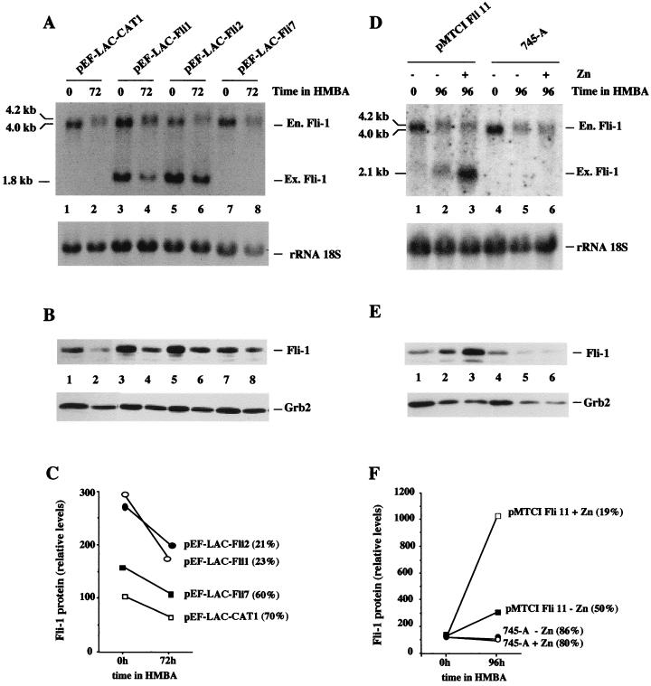
Deregulated overexpression of Fli-1 in 745-A cells inhibits their HMBA-induced erythroid differentiation. 745-A cells were stably transfected with empty vector pEF-LAC-CAT, Fli-1 constitutive expression vector pEF-LAC-Fli (A to C), or Fli-1 Zn-inducible Fli-1 expression vector pMTCI Fli (D to F), as indicated above the lanes, and individual clones were selected and amplified under appropriate antibiotic selection (G418 [1 mg/ml] for pEF-LAC-CAT or pEF-LAC-Fli and hygromycin [1 mg/ml] for pMTCI Fli). Individual clones or untransfected cells were grown for 3 (A to C) or 4 (D to F) days in the presence or absence of 5 mM HMBA with or without 200 μM ZnCl2 as indicated. The percentage of benzidine-positive differentiated cells was then determined, and total RNA and total protein cell lysates were prepared for Fli-1 RNA and protein analyses, respectively. (A and D) Northern blot sequentially hybridized with the Fli-1 probe (top) and the 18S rRNA probe (bottom). The positions of endogenous (En.) and exogenous (Ex.) Fli-1 transcripts are indicated on the right. (B and E) Results of Western blot analysis of Fli-1 proteins (top) and Grb2 proteins revealed on the same blot and taken as an internal standard (bottom). (C and F) Results of quantitative analyses of Fli-1 protein levels performed by densitometric tracing of the autoradiographs shown in panels B and E, respectively. Results are expressed as percentages of the Fli-1/Grb2 ratio determined for pEF-LAC-CAT1-transfected (C) and untransfected (F) 745-A cells, respectively. Numbers in parentheses indicate percentages of benzidine-positive cells.

