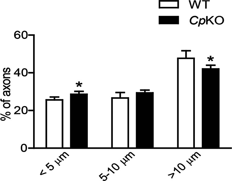
Figure 10.

Quantification of myelinated fiber diameters was done on Epon-embedded semithin sections stained with toluidine blue. The percent distribution is shown in three ranges. Note the slight shift from large- to small-diameter axons in Cp KO mice compared with WT mice. n = 3 per group. *p < 0.05.
