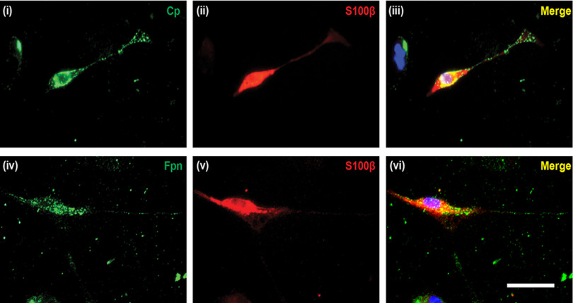
Figure 2.
Cell surface expression of Fpn and Cp in Schwann cells. Schwann cells were isolated from postnatal mouse (P5-P7 days old) sciatic nerve. Double immunofluorescence labeling with anti-Cp (i) or anti-Fpn (iv) and the Schwann cell marker anti-S100β (ii,v) antibodies demonstrated that Schwann cells express Cp and Fpn. iii, vi, Merged images, including DAPI staining, to visualize cell nuclei. Scale bar, 50 μm.

