Figure 5.
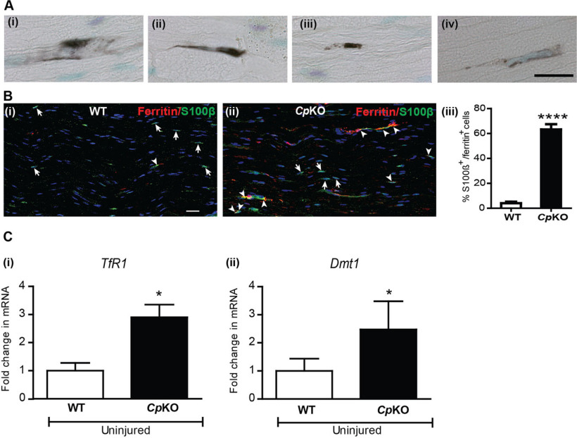
Iron accumulation in Schwann cells in Cp KO mice. Ai-Aiv, Iron histochemistry shows iron accumulation in cells that have a Schwann cell morphology in longitudinal sections of the sciatic nerve of Cp KO mice. Bi, Double immunofluorescence labeling of WT nerve for S100β (green) and ferrtin (red) and nuclear stain DAPI. Very few S100β+ Schwann cells express ferritin (arrowhead) in WT nerve. Single S100β+ cells (arrows). Bii, Cp KO nerve shows many S100β+ Schwann cells double-labeled for ferritin (arrowheads). Single S100β+ cells (arrows). Biii, Significant increase in the percentage of S100β+ Schwann cells double-labeled with ferritin in Cp KO nerves. n = 4 per group. ****p < 0.0001. Scale bars: Aiv, 20 μm; Bi, 15 μm. Ci, Cii, qPCR analysis of uninjured nerves from WT and Cp KO mice shows statistically significant increase in mRNA expression of iron importers TfR1 and Dmt1. n = 3 or 4 per group, each with 3 pooled nerves. *p = 0.03.

