Figure 6.

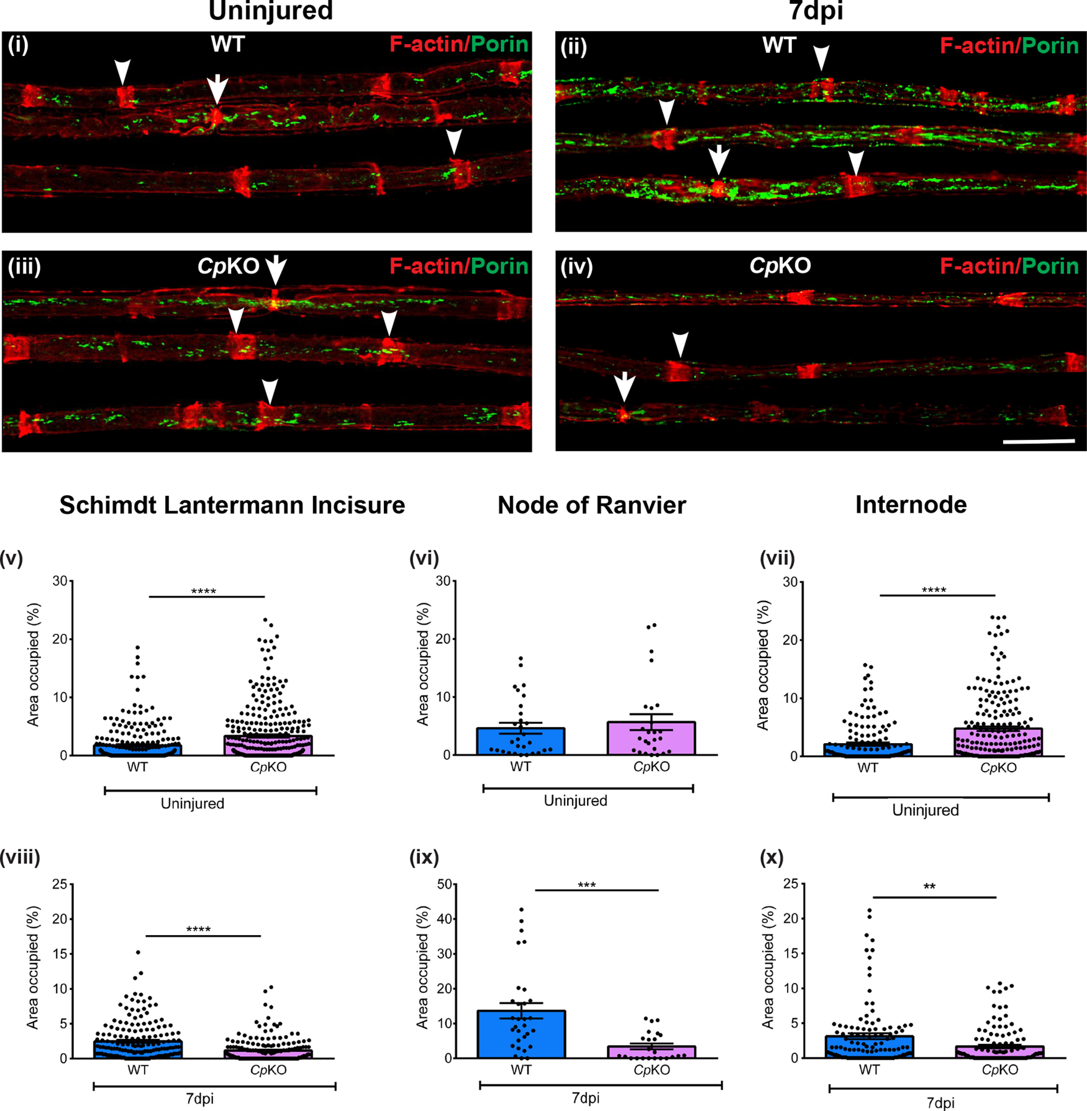
Changes in mitochondria in axons in Cp KO mice and effects of crush injury. Teased nerve fiber preparation of uninjured (i) and injured (ii) nerve fibers of WT mice. Note the marked increase in mitochondria in regenerating nerves of WT mice 7 d after crush injury (ii). Bottom panels, Uninjured (iii) and injured (iv) nerves of Cp KO mice. Note the marked reduction in mitochondria in regenerating nerves of Cp KO mice (iv) compared with WT mice (ii) 7 d after crush injury. Green represents mitochondrial profiles (porin staining). Red represents Schmidt-Lanterman incisures (arrowheads) and nodes of Ranvier (arrows) (rhodamine-phalloidin staining). v-x, Graphs represent area (%) occupied by mitochondria at the Schimdt-Lanterman incisures, nodes of Ranvier, and internode in teased axons from uninjured (v-vii) and injured (viii-x) sciatic nerves of WT and Cp KO mice. Uninjured teased fibers: Schmidt-Lanterman incisures, WT, n = 242, Cp KO mice, n = 301; nodes of Ranvier, WT mice, n = 25, Cp KO mice, n = 29; internode, WT, n = 158, Cp KO mice, n = 205 from 4 different animals of each genotype. Injured teased fibers: Schmidt-Lanterman incisures, WT mice, n = 174, Cp KO mice, n = 182; nodes of Ranvier, WT mice, n = 23, Cp KO mice, n = 30; internode, WT mice, n = 113, Cp KO mice, n = 120 from 3 different animals of each genotype. **p < 0.01. ***p < 0.001. ****p <0.0001. Scale bar, 20 µm.
