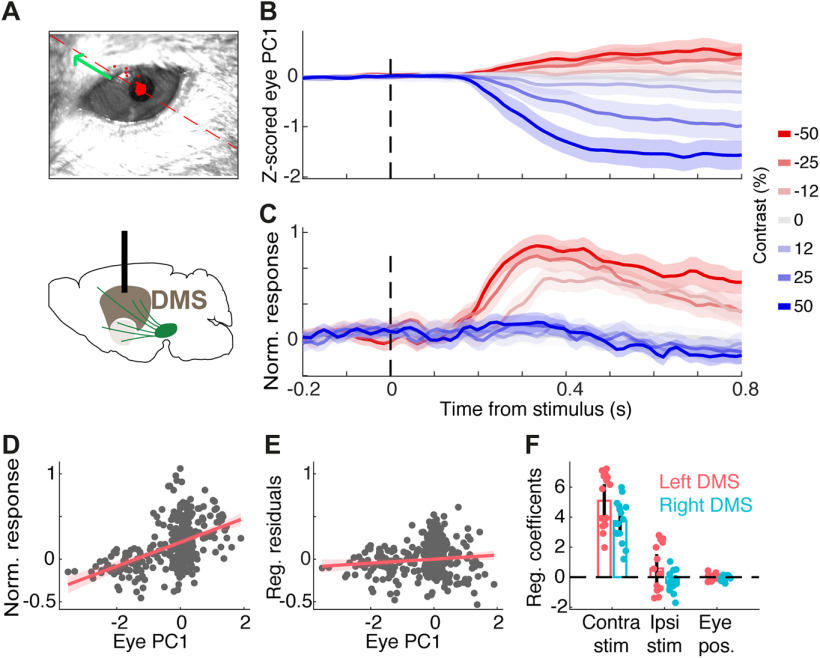
Figure 4.
DMS dopamine responses to contralateral stimuli cannot be explained by eye movements. A, Top: Example frame of the eye video. The red dashed line and green arrow indicates the positive direction of the first principal component (PC) of 2D eye position. All sessions with eye recordings were of the left eye. Bottom: Schematic of DMS dopamine recording. B, Z-scored first PC of pupil position in an example session. C, Dopamine signals recorded in the right DMS in the same session shown in B. D, The relationship between the first PC of pupil position and neural signals in the example session, before adjusting for the effect of stimulus contrast. Each dot indicates one trial. E, The relationship between the first PC of pupil position and neural signals after regressing out the confounding effect of stimulus contrast, indicating a negligible relationship between eye position and neural activity. F, The regression coefficients separately shown for sessions with left or right DMS dopamine recording in five mice. Each dot is one session and bars indicate averages across sessions. Coefficients of pupil position and ipsilateral stimuli were not significantly different from zero while coefficients of contralateral stimuli were significantly larger than zero (p = 0.96, p = 0.69, and p < 0.00001, respectively).

