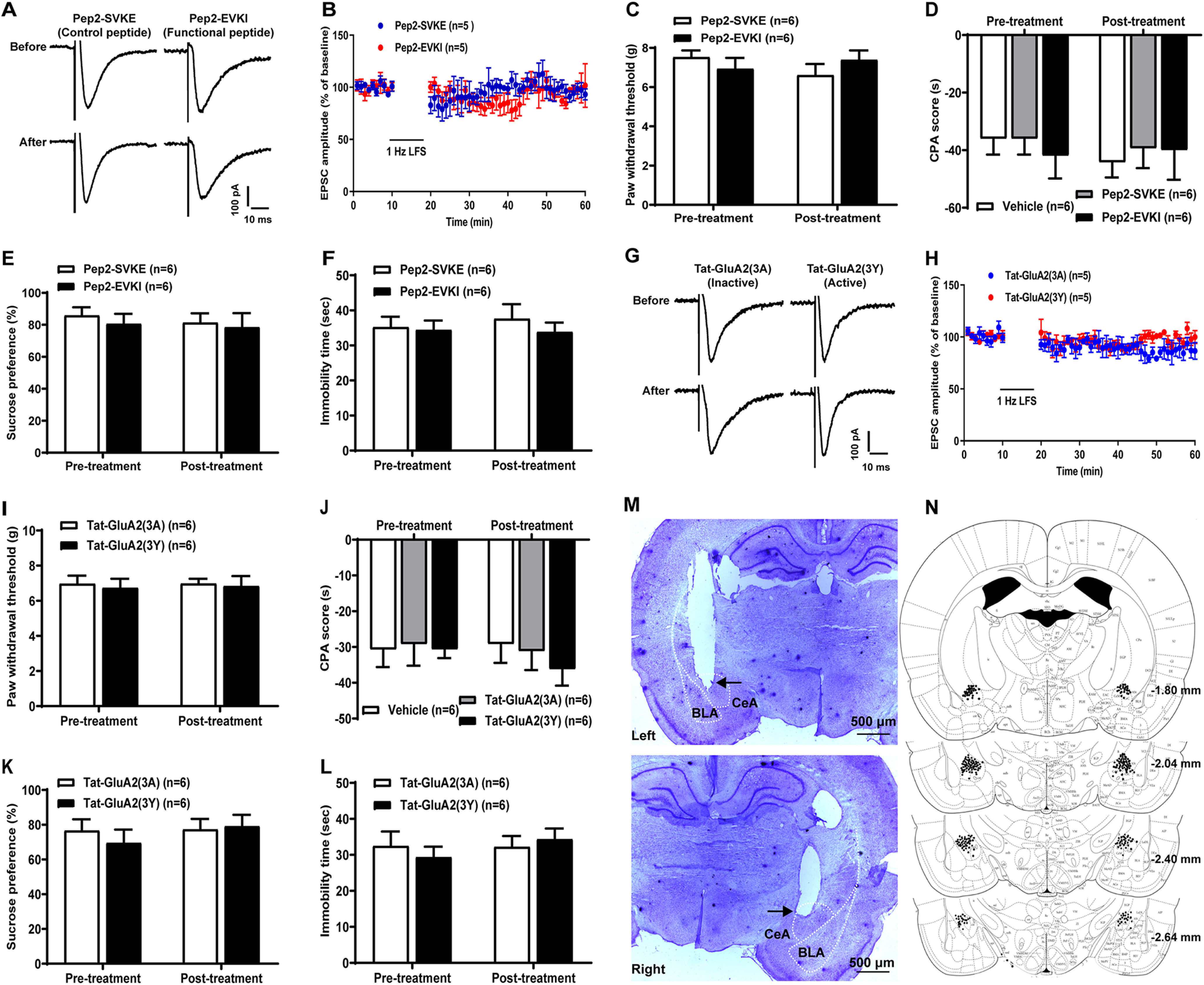
Figure 7.

Effects of microinjection of the peptides (pep2-EVKI/pep2-SVKE or Tat-GluA2(3Y)/Tat-GluA2(3A)) in bilateral CeA on LTD, pain sensitivity, and pain-related negative emotion in sham-operated rats. A–F, pep2-EVKI/pep2-SVKE. G-L, Tat-GluA2(3Y)/Tat-GluA2(3A). A, B, G, H, Effects of microinjection of the peptides on LTD of eEPSCs at the LA/BLA synapse. A, G, Representative traces of eEPSCs before and after LFS in pep2-EVKI/pep2-SVKE (A) and Tat-GluA2(3Y)/Tat-GluA2(3A) (G) groups. Calibration: 100 pA, 10 ms. B, H, Average eEPSC amplitudes (normalized to baseline) over the time course of the LFS protocol (480 pulses at 1 Hz with postsynaptic cell depolarized to −50 mV). C-F, I-L, Effects of microinjection of the peptides on pain sensitivity and pain-related negative emotion indicated by the PWT (C,I), CPA score (D,J), SPT (E,K), and immobility time in FST (F,L) in sham-operated rats (n = 6 rats per group). p > 0.05 (two-way ANOVA with Sidak's post hoc test). M, N, Histologically verified microinjection sites in the CeA. M, Examples of the Nissl-stained coronal section to illustrate the track of the microinjection cannula and the tip placement (as arrow shows) in left and right CeA. N, Representative four coronal sections through the rat amygdala are shown in sequence from anterior to posterior. Numbers in the right margin indicate millimeters posterior to the bregma. Filled circles in bilateral hemispheres represent the approximate positions of the cannula tips corresponding to some representative rats in the intra-CeA microinjection group. Diagrams are adapted from Paxinos and Watson (2014) and show coronal sections through bilateral hemispheres at different levels posterior to the bregma. Scale bar, 500 μm.
